

STD-11 UNIT-3 CHA-8
CELL : STRUCTURE AND FUNCTIONS
Flagella and Cilia
- They are fine hair like movable protoplasmic processes of the cells which are capable of producing a current in the fluid medium for locomotion and passage of substances.
- Flagella are longer ( 100-200 um ) but fewer.
- Only 1-4 flagella occur per cell, e.g. , many protists, motile algae, spermatozoa of animals, bryophytes and pteridophytes, choanocytes of sponges, gastrodermal cells of coelenterates, zoospores and gametes of thallophytes.
- Cilia are smaller (5–20 um) but are numerous.
- They occur in group ciliata of protista, flame cells of worms, larval bodies of many invertebrates, epithelium of respiratory tract, renal tubules, oviducal funnel, etc.
- Cilia present on the tracheal and bronchial epithelial cells are specialised to send back dust particles into the pharynx so that the lungs remain unharmed.
- However, cigarette smoking reduces / stops ciliary activity so that air borne dust particles pass into the lungs of smokers causing irrepairable harm.
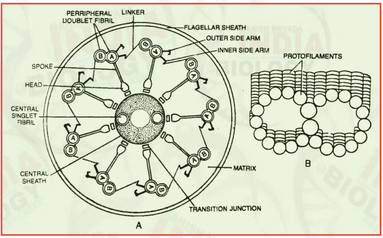
- Both cilia and flagella are structurally similar and possess similar parts- basal body, rootlets, basal plate and shaft.
- Basal Body or Kinetosome
- It is also called basal granule or blepharoplast
- Basal body occurs embedded in the outer part of the cytoplasm below the plasma membrane.
- It is like a microcylinder which has a structure similar to a centricle with nine triplet fibrils present on the periphery without a central fibril, though a hub of protein is present here.
- Only subfibre A is complete (having 13 protofilaments) while subfibres B and C are incomplete as they share some of their protofilaments.
- Rootlets
- They are striated fibrillar outgrowths which develop from the outer lower part of the basal body and are meant for providing support to the basal body.
- The rootlets are made of bundles of microfilaments.
- Basal Plate
- It is an area of high density which lies above the basal body at the level of the plasma membrane.
- In the region of basal plate, one sub-fiber of each peripheral fibril disappears.
- The central fibrils develop in this area.
- Shaft.
- It is the hair - like projecting part of flagellum or cilium.
- The length is 5 - 20 um in case of cilium and 100-200 um in case of flagellum.
- The shaft is covered on the outside by a sheath which is the extension of plasma membrane.
- In whiplash Flagellum, the sheath is smooth.
- In tinsel flagellum, the sheath contains a number of thick hairy outgrowths called flimmers.
- Internally, it contains a semifluid matrix having an axoneme of 9 peripheral doublet fibrils and 2 central singlet fibrils.
- This arrangement is called 9 + 2 or 11- stranded.
- However 9 + 1 (e.g., flatworm) and 9 +0 (e.g., eel, Asian Horseshoe Crab) arrangements have also been observed.
- The two central singlet fibers are covered by a proteinaceous central sheath.
- They are connected by a double bridge.
- Each peripheral fibril consists of two microtubules or sub-fibers B and A.
- The sub-fiber A is slightly narrower. It bears two bent arms, the outer one having a hook.
- They are about 15 nm long and made up of protein dynein with ATP - ase activity.
- Such activity is also present in central fibrils.
- Movement of flagella or cilia occurs due to sliding motion in which dynein arm establishes temporary connection with subtubule B of adjacent doublet fiber.
- The pe ripheral doublet fibrils as well as central singlet fibrils are made up of tubulin.
- Each sub-fiber or central singlet fibril contains thirteen protofilaments. The peripheral doublet fibrils are interconnected by A - B linkers of protein nexin between B - subfiber of one and inner side arm of A - subfiber of adjacent fibril.
- Each of their A sub-fibers sends a radial proteinaceous column to the center. It is called spoke.
- The spokes are broader internally to form heads or knobs.
- Head is connected to central proteinaceous sheath through transition junction.

- The cilia and Nagella move by sliding of the doublet fibrils against one another.
- Energy is provided by ATP.
- Flagella perform independent undulatory movements while cilia show rowing type of sweeping motion either simultaneously (isochronic or synchronous) or one after the other (metachronic).
- In a flagellum, sev eral symmetrical undulatory waves pass from base to the tip.
- This pushes the cell along. Undulations passing from tip to base pull the cell through water.
- In tinsel flagellum having a number of flimmers, the undulatory wave moving down from base to tip also pulls the cell along instead of pushing it.
- There is always a power stroke and a recovery or return stroke.
- The power stroke is able to move the fluid with a jerk in the direction of the stroke.
- The cell moves in the opposite direction, if it is motile.
- The recovery or return stroke is slow and without much force.
- Therefore, it does not cause much disturbance in the fluid medium.
- Rate of cili ary and flagellar movements is 10–40 strokes per second.
- Flagellate Monas stigmatica swims at the rate of 260 pm or 40 cell length/sec.
- It has the maximum speed per body length.
- Paramoecium caudatum has a speed of 1500 pm or 12 cell lengths sec.
- They help in locomotion in flagellate and ciliated organisms.
- They create current for obtaining food from aquatic medium.
- In some protists and animals , the organelles take part in capturing food .
- The canal system of porifers operates with the help of flagella present in their collar cells or choanocytes.
- In coelenterates , they circulate food in the gastrovascular cavity. In tunicates and lancelets, the cilia help in movement of food and its egestion.
- In aquatic organisms cilia create currents in water for renewal of oxygen supply and quick dif fusion of carbon dioxide.
- In land animals the cilia of the respiratory tract help in eliminating dust particles in the incom ing air.
- Internal transport of several organs is performed by cilia , e.g. passage of eggs in oviduct , passage of excretory substances in the kidneys , etc.
- Being protoplasmic structures they can function as sensory organs.
- Their tips secrete sticky substance to help in conjugation and fusion of gametes.
- In certain protistans, cilia fuse to form undulating membrane.
- Cilia and flagella show sensitivity to changes in light, temperature and contact.
- Ciliated larvae take part in dispersal of the species.
========================================


Please do not enter any spam link or word in the comment box